GRIK2
Protein-coding gene in the species Homo sapiens
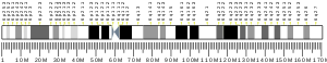
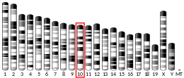
| GRIK2 | |||||||||||||||||||||||||||||||||||||||||||||||||||
|---|---|---|---|---|---|---|---|---|---|---|---|---|---|---|---|---|---|---|---|---|---|---|---|---|---|---|---|---|---|---|---|---|---|---|---|---|---|---|---|---|---|---|---|---|---|---|---|---|---|---|---|
 | |||||||||||||||||||||||||||||||||||||||||||||||||||
| Identifiers | |||||||||||||||||||||||||||||||||||||||||||||||||||
| Aliases | GRIK2, EAA4, GLR6, GLUK6, GLUR6, GluK2, MRT6, glutamate ionotropic receptor kainate type subunit 2, NEDLAS | ||||||||||||||||||||||||||||||||||||||||||||||||||
| External IDs | OMIM: 138244 MGI: 95815 HomoloGene: 40717 GeneCards: GRIK2 | ||||||||||||||||||||||||||||||||||||||||||||||||||
| |||||||||||||||||||||||||||||||||||||||||||||||||||
| |||||||||||||||||||||||||||||||||||||||||||||||||||
| |||||||||||||||||||||||||||||||||||||||||||||||||||
| |||||||||||||||||||||||||||||||||||||||||||||||||||
| |||||||||||||||||||||||||||||||||||||||||||||||||||
| Wikidata | |||||||||||||||||||||||||||||||||||||||||||||||||||
| |||||||||||||||||||||||||||||||||||||||||||||||||||


Glutamate ionotropic receptor kainate type subunit 2, also known as ionotropic glutamate receptor 6 or GluR6, is a protein that in humans is encoded by the GRIK2 (or GLUR6) gene.[5][6][7]
Function
This gene encodes a subunit of a kainate glutamate receptor. This receptor may have a role in synaptic plasticity, learning, and memory. It also may be involved in the transmission of visual information from the retina to the hypothalamus. The structure and function of the encoded protein is influenced by RNA editing. Alternatively spliced transcript variants encoding distinct isoforms have been described for this gene.[7] It has been discovered that this is a key protein, which enables mammals to feel cold sensations.[8]
Clinical significance
Homozygosity for a GRIK2 deletion-inversion mutation is associated with non-syndromic autosomal recessive mental retardation.[9]
Interactions
GRIK2 has been shown to interact with:
- DLG1,[10][11]
- DLG4,[10][11][12]
- GRID2,[13]
- GRIK5,[14][15]
- GRIP1,[12][16]
- PICK1[12] and
- SDCBP.[12][16]
RNA Editing
Pre-mRNA for several neurotransmitter receptors and ion channels are substrates for ADARs, including AMPA receptor subunits (GluR2, GluR3, GluR4) and kainate receptor subunits (GluR5, GluR6). Glutamate-gated ion channels are made up of four subunits per channel, with each subunit contributing to the pore loop structure. The pore loop structure is similar to that found in K+ channels (e.g. the human Kv1.1 channel, whose pre-mRNA is also subject to A to I RNA editing).[17][18] The diversity of ionotropic glutamate receptor subunits, as well as RNA splicing, is determined by RNA editing events of the individual subunits, explaining their extremely high diversity.
Type
The type of RNA editing that occurs in the pre-mRNA of GluR6 is Adenosine to Inosine (A to I) editing. [19]
A to I RNA editing is catalyzed by a family of adenosine deaminases acting on RNA (ADARs) that specifically recognize adenosines within double-stranded regions of pre-mRNAs and deaminate them to inosine. Inosines are recognised as guanosine by the cell's translational machinery. There are three members of the ADAR family ADARs 1–3 with ADAR1 and ADAR2 being the only enzymatically active members. ADAR1 and ADAR2 are widely expressed in tissues, while ADAR3 is restricted to the brain, where it is though tot have a regulatory role. The double-stranded regions of RNA are formed by base-pairing between residues close to region of the editing site, with residues usually in a neighboring intron, though they can occasionally be located in an exonic sequence. The region that forms base pairs with the editing region is known as an Editing Complementary Sequence (ECS).
ADARs bind interact directly with the dsRNA substrate via their double-stranded RNA binding domains. If an editing site occurs within a coding sequence, the result could be a codon change. This can lead to translation of a protein isoform due to a change in its primary protein structure. Therefore, editing can also alter protein function. A to I editing occurs in a noncoding RNA sequences such as introns, untranslated regions (UTRs), LINEs, and SINEs (especially Alu repeats). The function of A to I editing in these regions is thought to involve creation of splice sites and retention of RNAs in the nucleus amongst others.
Location
The pre-mRNA of GLUR6 is edited at amino acid positions 567, 571, and 621. The Q/R position, which gets its name as editing results in an codon change from a glutamine (Q) codon (CAG) to an arginine (R) codon (CGG), is located in the "pore loop" of the second membrane domain (M2). The Q/R site of GluR6 pre-mRNA occurs in an asymmetrical loop of three exonic and four intronic nucleotides. The Q/R editing site is also observed in GluR2 and GluR5. The Q/R site is located in a homologous position in GluR2 and in GluR6.[20]
GluR-6 is also edited at I/V and Y/C sites, which are found in the first membrane domain (M1). At the I/V site, editing results in a codon change from (ATT) isoleucine (I) to (GTT) valine (V), while at the Y/C site, the codon change is from (TAC) tyrosine (Y) to (TGC) cysteine (C).[21]
The RNAfold program characterised a putative double-stranded RNA (dsRNA) conformation around the Q/R site of the GluR-6 pre-mRNA. This sequence is necessary for editing at the site to occur. The possible editing complementary sequence was observed from transcript analysis to be 1.9 kb downstream from the editing site within intron 12.[20] The ECS for the editing sites in M1 has yet to be identified but it is likely to occur at a considerable distance from the editing sites.[22]
Regulation
Editing of the Q/R site in GluR6 pre-mRNA has been demonstrated to be developmentally regulated in rats, ranging from 0% in rat embryo to 80% at birth. This is different from the AMPA receptor subunit GluR2, which is nearly 100% edited and is not developmentally regulated.[21] Significant amounts of both edited and non-edited forms of GluR6 transcripts are found in the adult brain. The receptor is 90% edited in all grey matter structures, while in white matter, the receptor is edited in just 10% of cases. Frequency increases from 0% in rat embryo to 85% in adult rat.
Consequences
Structure
The primary GluR6 transcripts can be edited in up to three positions. Editing at each of the three positions affects Ca2+ permeability of the channel.[23]
Function
Editing plays a role in the electrophysiology of the channel. Editing at the Q/R site has been deemed to be nonessential in GluR6.[24] It has been reported that the unedited version of GluR6 functions in the regulation of synaptic plasticity. The edited version is thought to inhibit synaptic plasticity and reduce seizure susceptibility.[23] Mice lacking the Q/R site exhibit increased long term potentiation and are more susceptible to kainate induced seizures. The number of seizures is inversely correlated with the amount of RNA editing. Human GluR6 pre-mRNA editing is increased during seizures, possibly as an adaptive mechanism.[25][26]
Up to 8 different protein isoforms can occur as a result of different combinations of editing at the three sites, giving rise to receptor variants with differing kinetics. The effect of Q/R site editing on calcium permeability appears to be dependent on editing of the I/V and Y/C sites. When both sites in TM1 (I/V and Y/C) are edited, Q/R site editing is required for calcium permeability. On the contrary, when neither the I/V nor the Y/C site is edited, receptors demonstrate high calcium permeability regardless of Q/R site editing. The co-assembly of these two isoforms generate receptors with reduced calcium permeability.[23]
RNA editing of the Q/R site can affect inhibition of the channel by membrane fatty acids such as arachidonic acid and docosahexaenoic acid[27] For Kainate receptors with only edited isforms, these are strongly inhibited by these fatty acids, however inclusion of just one non-edited subunit is enough to abolish this effect.[27]
Dysregulation
Kainate-induced seizures in mice are used as a model of temporal lobe epilepsy in humans. Despite mice deficient in editing at the Q/R site of GluR6 showing increased seizure susceptibility, tissue analysis of human epilepsy patients did not show reduced editing at this site.[24][28][29][30]
See also
References
- ^ a b c GRCh38: Ensembl release 89: ENSG00000164418 – Ensembl, May 2017
- ^ a b c GRCm38: Ensembl release 89: ENSMUSG00000056073 – Ensembl, May 2017
- ^ "Human PubMed Reference:". National Center for Biotechnology Information, U.S. National Library of Medicine.
- ^ "Mouse PubMed Reference:". National Center for Biotechnology Information, U.S. National Library of Medicine.
- ^ HGNC. "Symbol Report: GRIK2". Retrieved 29 December 2017.
- ^ Paschen W, Blackstone CD, Huganir RL, Ross CA (Aug 1994). "Human GluR6 kainate receptor (GRIK2): molecular cloning, expression, polymorphism, and chromosomal assignment". Genomics. 20 (3): 435–40. doi:10.1006/geno.1994.1198. PMID 8034316.
- ^ a b "Entrez Gene: GRIK2 glutamate receptor, ionotropic, kainate 2".
- ^ Sherburne, Morgan (11 March 2024). "Unlocking the Chill: Protein Behind Cold Sensation Found".
- ^ Motazacker MM, Rost BR, Hucho T, Garshasbi M, Kahrizi K, Ullmann R, Abedini SS, Nieh SE, Amini SH, Goswami C, Tzschach A, Jensen LR, Schmitz D, Ropers HH, Najmabadi H, Kuss AW (October 2007). "A defect in the ionotropic glutamate receptor 6 gene (GRIK2) is associated with autosomal recessive mental retardation". Am. J. Hum. Genet. 81 (4): 792–8. doi:10.1086/521275. PMC 2227928. PMID 17847003.
- ^ a b Mehta S, Wu H, Garner CC, Marshall J (May 2001). "Molecular mechanisms regulating the differential association of kainate receptor subunits with SAP90/PSD-95 and SAP97". J. Biol. Chem. 276 (19): 16092–9. doi:10.1074/jbc.M100643200. PMID 11279111.
- ^ a b Garcia EP, Mehta S, Blair LA, Wells DG, Shang J, Fukushima T, Fallon JR, Garner CC, Marshall J (Oct 1998). "SAP90 binds and clusters kainate receptors causing incomplete desensitization". Neuron. 21 (4): 727–39. doi:10.1016/s0896-6273(00)80590-5. PMID 9808460. S2CID 18723258.
- ^ a b c d Hirbec H, Francis JC, Lauri SE, Braithwaite SP, Coussen F, Mulle C, Dev KK, Coutinho V, Meyer G, Isaac JT, Collingridge GL, Henley JM, Couthino V (Feb 2003). "Rapid and differential regulation of AMPA and kainate receptors at hippocampal mossy fibre synapses by PICK1 and GRIP". Neuron. 37 (4): 625–38. doi:10.1016/s0896-6273(02)01191-1. PMC 3314502. PMID 12597860.
- ^ Kohda K, Kamiya Y, Matsuda S, Kato K, Umemori H, Yuzaki M (Jan 2003). "Heteromer formation of delta2 glutamate receptors with AMPA or kainate receptors". Brain Res. Mol. Brain Res. 110 (1): 27–37. doi:10.1016/s0169-328x(02)00561-2. PMID 12573530.
- ^ Wenthold RJ, Trumpy VA, Zhu WS, Petralia RS (Jan 1994). "Biochemical and assembly properties of GluR6 and KA2, two members of the kainate receptor family, determined with subunit-specific antibodies". J. Biol. Chem. 269 (2): 1332–9. doi:10.1016/S0021-9258(17)42262-9. PMID 8288598.
- ^ Ripellino JA, Neve RL, Howe JR (Jan 1998). "Expression and heteromeric interactions of non-N-methyl-D-aspartate glutamate receptor subunits in the developing and adult cerebellum". Neuroscience. 82 (2): 485–97. doi:10.1016/s0306-4522(97)00296-0. PMID 9466455. S2CID 23219004.
- ^ a b Hirbec H, Perestenko O, Nishimune A, Meyer G, Nakanishi S, Henley JM, Dev KK (May 2002). "The PDZ proteins PICK1, GRIP, and syntenin bind multiple glutamate receptor subtypes. Analysis of PDZ binding motifs". J. Biol. Chem. 277 (18): 15221–4. doi:10.1074/jbc.C200112200. hdl:2262/89271. PMID 11891216.
- ^ Seeburg PH, Single F, Kuner T, Higuchi M, Sprengel R (July 2001). "Genetic manipulation of key determinants of ion flow in glutamate receptor channels in the mouse". Brain Res. 907 (1–2): 233–43. doi:10.1016/S0006-8993(01)02445-3. PMID 11430906. S2CID 11969068.
- ^ Bhalla T, Rosenthal JJ, Holmgren M, Reenan R (October 2004). "Control of human potassium channel inactivation by editing of a small mRNA hairpin". Nat. Struct. Mol. Biol. 11 (10): 950–6. doi:10.1038/nsmb825. PMID 15361858. S2CID 34081059.
- ^ 52. Seeburg PH, Higuchi M, Sprengel R. Brain Res Brain Res Rev. 1998;26:217–29.
- ^ a b Sommer B, Köhler M, Sprengel R, Seeburg PH (October 1991). "RNA editing in brain controls a determinant of ion flow in glutamate-gated channels". Cell. 67 (1): 11–9. doi:10.1016/0092-8674(91)90568-J. PMID 1717158. S2CID 22029384.
- ^ a b Bernard A, Khrestchatisky M (May 1994). "Assessing the extent of RNA editing in the TMII regions of GluR5 and GluR6 kainate receptors during rat brain development". J. Neurochem. 62 (5): 2057–60. doi:10.1046/j.1471-4159.1994.62052057.x. PMID 7512622. S2CID 27091741.
- ^ Niswender CM (September 1998). "Recent advances in mammalian RNA editing". Cell. Mol. Life Sci. 54 (9): 946–64. doi:10.1007/s000180050225. PMID 9791538. S2CID 20556833.
- ^ a b c Köhler M, Burnashev N, Sakmann B, Seeburg PH (March 1993). "Determinants of Ca2+ permeability in both TM1 and TM2 of high affinity kainate receptor channels: diversity by RNA editing". Neuron. 10 (3): 491–500. doi:10.1016/0896-6273(93)90336-P. PMID 7681676. S2CID 39976579.
- ^ a b Vissel B, Royle GA, Christie BR, Schiffer HH, Ghetti A, Tritto T, Perez-Otano I, Radcliffe RA, Seamans J, Sejnowski T, Wehner JM, Collins AC, O'Gorman S, Heinemann SF (January 2001). "The role of RNA editing of kainate receptors in synaptic plasticity and seizures". Neuron. 29 (1): 217–27. doi:10.1016/S0896-6273(01)00192-1. PMID 11182093. S2CID 7976952.
- ^ Bernard A, Ferhat L, Dessi F, Charton G, Represa A, Ben-Ari Y, Khrestchatisky M (February 1999). "Q/R editing of the rat GluR5 and GluR6 kainate receptors in vivo and in vitro: evidence for independent developmental, pathological and cellular regulation". Eur. J. Neurosci. 11 (2): 604–16. doi:10.1046/j.1460-9568.1999.00479.x. PMID 10051761. S2CID 7866926.
- ^ Grigorenko EV, Bell WL, Glazier S, Pons T, Deadwyler S (July 1998). "Editing status at the Q/R site of the GluR2 and GluR6 glutamate receptor subunits in the surgically excised hippocampus of patients with refractory epilepsy". NeuroReport. 9 (10): 2219–24. doi:10.1097/00001756-199807130-00013. PMID 9694203. S2CID 28692872.
- ^ a b Wilding TJ, Fulling E, Zhou Y, Huettner JE (July 2008). "Amino acid substitutions in the pore helix of GluR6 control inhibition by membrane fatty acids". J. Gen. Physiol. 132 (1): 85–99. doi:10.1085/jgp.200810009. PMC 2442176. PMID 18562501.
- ^ Nadler JV (November 1981). "Minireview. Kainic acid as a tool for the study of temporal lobe epilepsy". Life Sci. 29 (20): 2031–42. doi:10.1016/0024-3205(81)90659-7. PMID 7031398.
- ^ Ben-Ari Y (February 1985). "Limbic seizure and brain damage produced by kainic acid: mechanisms and relevance to human temporal lobe epilepsy". Neuroscience. 14 (2): 375–403. doi:10.1016/0306-4522(85)90299-4. PMID 2859548. S2CID 33597110.
- ^ Kortenbruck G, Berger E, Speckmann EJ, Musshoff U (June 2001). "RNA editing at the Q/R site for the glutamate receptor subunits GLUR2, GLUR5, and GLUR6 in hippocampus and temporal cortex from epileptic patients". Neurobiol. Dis. 8 (3): 459–68. doi:10.1006/nbdi.2001.0394. PMID 11442354. S2CID 33605674.
Further reading
- Seeburg PH, Higuchi M, Sprengel R (1998). "RNA editing of brain glutamate receptor channels: mechanism and physiology". Brain Res. Brain Res. Rev. 26 (2–3): 217–29. doi:10.1016/S0165-0173(97)00062-3. PMID 9651532. S2CID 12147763.
- Paschen W, Hedreen JC, Ross CA (1994). "RNA editing of the glutamate receptor subunits GluR2 and GluR6 in human brain tissue". J. Neurochem. 63 (5): 1596–602. doi:10.1046/j.1471-4159.1994.63051596.x. PMID 7523595. S2CID 25226376.
- Hoo KH, Nutt SL, Fletcher EJ, Elliott CE, Korczak B, Deverill RM, Rampersad V, Fantaske RP, Kamboj RK (1995). "Functional expression and pharmacological characterization of the human EAA4 (GluR6) glutamate receptor: a kainate selective channel subunit". Recept. Channels. 2 (4): 327–37. PMID 7536611.
- Sander T, Janz D, Ramel C, Ross CA, Paschen W, Hildmann T, Wienker TF, Bianchi A, Bauer G, Sailer U (1995). "Refinement of map position of the human GluR6 kainate receptor gene (GRIK2) and lack of association and linkage with idiopathic generalized epilepsies". Neurology. 45 (9): 1713–20. doi:10.1212/wnl.45.9.1713. PMID 7675232. S2CID 24350236.
- Nutt SL, Kamboj RK (1995). "RNA editing of human kainate receptor subunits". NeuroReport. 5 (18): 2625–9. doi:10.1097/00001756-199412000-00055. PMID 7696618.
- Raymond LA, Blackstone CD, Huganir RL (1993). "Phosphorylation and modulation of recombinant GluR6 glutamate receptors by cAMP-dependent protein kinase". Nature. 361 (6413): 637–41. Bibcode:1993Natur.361..637R. doi:10.1038/361637a0. PMID 8094892. S2CID 4339168.
- Roche KW, Raymond LA, Blackstone C, Huganir RL (1994). "Transmembrane topology of the glutamate receptor subunit GluR6". J. Biol. Chem. 269 (16): 11679–82. doi:10.1016/S0021-9258(17)32623-6. PMID 8163463.
- Taverna FA, Wang LY, MacDonald JF, Hampson DR (1994). "A transmembrane model for an ionotropic glutamate receptor predicted on the basis of the location of asparagine-linked oligosaccharides". J. Biol. Chem. 269 (19): 14159–64. doi:10.1016/S0021-9258(17)36768-6. PMID 8188697.
- Wenthold RJ, Trumpy VA, Zhu WS, Petralia RS (1994). "Biochemical and assembly properties of GluR6 and KA2, two members of the kainate receptor family, determined with subunit-specific antibodies". J. Biol. Chem. 269 (2): 1332–9. doi:10.1016/S0021-9258(17)42262-9. PMID 8288598.
- Pickering DS, Taverna FA, Salter MW, Hampson DR (1996). "Palmitoylation of the GluR6 kainate receptor". Proc. Natl. Acad. Sci. U.S.A. 92 (26): 12090–4. doi:10.1073/pnas.92.26.12090. PMC 40302. PMID 8618850.
- Bonaldo MF, Lennon G, Soares MB (1997). "Normalization and subtraction: two approaches to facilitate gene discovery". Genome Res. 6 (9): 791–806. doi:10.1101/gr.6.9.791. PMID 8889548.
- Porter RH, Eastwood SL, Harrison PJ (1997). "Distribution of kainate receptor subunit mRNAs in human hippocampus, neocortex and cerebellum, and bilateral reduction of hippocampal GluR6 and KA2 transcripts in schizophrenia". Brain Res. 751 (2): 217–31. doi:10.1016/S0006-8993(96)01404-7. PMID 9099808. S2CID 9796632.
- Rubinsztein DC, Leggo J, Chiano M, Dodge A, Norbury G, Rosser E, Craufurd D (1997). "Genotypes at the GluR6 kainate receptor locus are associated with variation in the age of onset of Huntington disease". Proc. Natl. Acad. Sci. U.S.A. 94 (8): 3872–6. Bibcode:1997PNAS...94.3872R. doi:10.1073/pnas.94.8.3872. PMC 20534. PMID 9108071.
- Ripellino JA, Neve RL, Howe JR (1998). "Expression and heteromeric interactions of non-N-methyl-D-aspartate glutamate receptor subunits in the developing and adult cerebellum". Neuroscience. 82 (2): 485–97. doi:10.1016/S0306-4522(97)00296-0. PMID 9466455. S2CID 23219004.
- Garcia EP, Mehta S, Blair LA, Wells DG, Shang J, Fukushima T, Fallon JR, Garner CC, Marshall J (1998). "SAP90 binds and clusters kainate receptors causing incomplete desensitization". Neuron. 21 (4): 727–39. doi:10.1016/S0896-6273(00)80590-5. PMID 9808460. S2CID 18723258.
- Leuschner WD, Hoch W (1999). "Subtype-specific assembly of alpha-amino-3-hydroxy-5-methyl-4-isoxazole propionic acid receptor subunits is mediated by their n-terminal domains". J. Biol. Chem. 274 (24): 16907–16. doi:10.1074/jbc.274.24.16907. PMID 10358037.
- Smith HJ (2001). "The introduction of MR in the Nordic countries with special reference to Norway: central control versus local initiatives". Journal of Magnetic Resonance Imaging. 13 (4): 639–44. doi:10.1002/jmri.1090. PMID 11276111. S2CID 25114658.
- Mehta S, Wu H, Garner CC, Marshall J (2001). "Molecular mechanisms regulating the differential association of kainate receptor subunits with SAP90/PSD-95 and SAP97". J. Biol. Chem. 276 (19): 16092–9. doi:10.1074/jbc.M100643200. PMID 11279111.
External links
- GRIK2+protein,+human at the U.S. National Library of Medicine Medical Subject Headings (MeSH)
This article incorporates text from the United States National Library of Medicine, which is in the public domain.
- v
- t
- e
PDB gallery
-
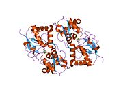
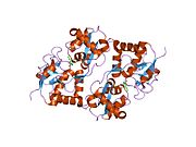
 1s50: X-ray structure of the GluR6 ligand binding core (S1S2A) in complex with glutamate at 1.65 A resolution
 -
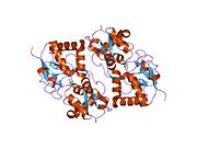
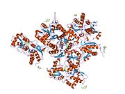
 1s7y: Crystal structure of the GluR6 ligand binding core in complex with glutamate at 1.75 A resolution orthorhombic form
 -
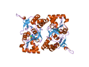
 1s9t: Crystal structure of the GLUR6 ligand binding core in complex with quisqualate at 1.8A resolution
 -
 1sd3: Crystal structure of the GLUR6 ligand binding core in complex with 2S,4R-4-methylglutamate at 1.8 Angstrom resolution
 -
 1tt1: CRYSTAL STRUCTURE OF THE GLUR6 LIGAND BINDING CORE IN COMPLEX WITH KAINATE 1.93 A RESOLUTION
 -
 1yae: Structure of the Kainate Receptor Subunit GluR6 Agonist Binding Domain Complexed with Domoic Acid
 -
 2i0b: Crystal structure of the GluR6 ligand binding core ELKQ mutant dimer at 1.96 Angstroms Resolution
 -
 2i0c: Crystal structure of the GluR6 ligand binding core dimer crosslinked by disulfide bonds between Y490C and L752C at 2.25 Angstroms Resolution


